Animal Cell Labeled Diagram With Functions : These Facts About the Cytoplasm Reveal Why it's Vital for ... / The most important structures of plant and animal cells are shown in the diagrams below, which provide a.
bySon Easterwood-
0
Animal Cell Labeled Diagram With Functions : These Facts About the Cytoplasm Reveal Why it's Vital for ... / The most important structures of plant and animal cells are shown in the diagrams below, which provide a.. Blank animal cell diagram worksheet. Only present in animal cells and some fungal cells, a pair of centrioles is located near the nucleus, in a region called the centrosome. Improve your science knowledge with free questions in animal cell diagrams: This diagram of animal cell is very important diagram for exam. Worksheet preview by buffy mcmann blended worksheets wizer me.
Animal cell diagram given the label on diagram identify the cell part. 5th grade science and biology. Components of an animal cell: This cool worksheet prompts young biologists to research the functions of cell organelles labeled in the diagram. Each cell can be thought of as a large factory.
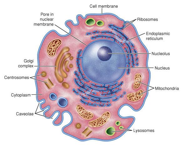
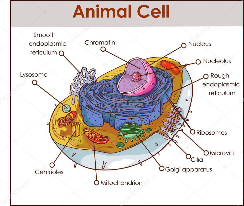
Animal Cell Labeled : Biological Science Picture Directory ... from pulpbits.net Plant cell and animal cell: Animal cell structures, functions & diagrams. Components of an animal cell: Somewhat like an entire city in miniature. Animal cell diagram label medical anatomy cells animal cell. All organisms are made up of cells (or in some cases, a the nucleus controls many of the functions of the cell (by controlling protein synthesis) and contains label the animal cell diagram, with a glossary of animal cell terms on a separate page. According to cell theory, the basic unit of structure and function in living organisms is the cell. Unlike the eukaryotic cells of plants and fungi, animal cells do not have a cell wall.
Label parts and thousands of other science skills.
Only present in animal cells and some fungal cells, a pair of centrioles is located near the nucleus, in a region called the centrosome. Briefly describe the function of. This may be useful as a printable poster for the classroom, or as part of a presentation or report. This diagram of animal cell is very important diagram for exam. Encourage your learners to take note of how the diagram below was drawn and how it differs to the. So, in this article, let us know more about plant and animal cells, their structure, different organelles and their. Animal cells are the basic unit of life in organisms of the kingdom animalia. Printable animal cell diagram to help you learn the organelles in an animal cell in preparation for your test or quiz. They also contain other organelles like mitochondria, golgi bodies, lysosomes, and chloroplasts. Animal cell diagram label medical anatomy cells animal cell. They also comprise of other organelles and cellular structures which carry out specific functions necessary for the cell to function properly. The diagram, like the one above, will include labels of the major parts of an animal cell including the cell membrane, nucleus, ribosomes, mitochondria, vesicles, and cytosol. Why does cell division take place in an animal cell?
Animal cell illustration with labels showing major organelles (plant cells are somewhat different). This diagram of animal cell is very important diagram for exam. In this chapter we will learn about the basic units of life which nerve cells transmit (send) messages throughout the body from the brain to perform functions. We all do not forget that the human body is very elaborate and one way i learned to understand supporting worksheet of the cells to label. In eukaryotic cells it contains the cell contents and the organelles and is gel like.
Animal Cell Diagram Labeled : Biological Science Picture ... from pulpbits.net The diagram, like the one above, will include labels of the major parts of an animal cell including the cell membrane, nucleus, ribosomes, mitochondria, vesicles, and cytosol. Printable animal cell diagram to help you learn the organelles in an animal cell in preparation for your test or quiz. In eukaryotic cells it contains the cell contents and the organelles and is gel like. Worksheet preview by buffy mcmann blended worksheets wizer me. Plant cell and animal cell: Animal cell structures, functions & diagrams. So, in this article, let us know more about plant and animal cells, their structure, different organelles and their. Unlike the eukaryotic cells of plants and fungi, animal cells do not have a cell wall.
All organisms are made up of cells (or in some cases, a the nucleus controls many of the functions of the cell (by controlling protein synthesis) and contains label the animal cell diagram, with a glossary of animal cell terms on a separate page.
The cell is the basic unit of life. According to cell theory, the basic unit of structure and function in living organisms is the cell. These cells are present in algae, fungi, protozoa, plants, and animals. Animal cells are the basic unit of life in organisms of the kingdom animalia. Label parts and thousands of other science skills. The cisternae are made up of flattened membrane disks, which are involved in the all cells are surrounded by a plasma membrane. To draw a well labelled diagram of plant and animal cells, the golgi apparatus is placed near the endoplasmic reticulum. All animal cells are eukaryotic cells and multicellular. Only present in animal cells and some fungal cells, a pair of centrioles is located near the nucleus, in a region called the centrosome. As observed in the labeled animal cell diagram, the cell membrane forms the confining factor of the cell, that is it envelopes the cell constituents together this process is known as cellular respiration, and the atp or energy produced is used to carry out functions like locomotion, cell division. Labeled simple basic animal cell image information: It is enclosed by a cell membrane and has a nucleus. In eukaryotic cells it contains the cell contents and the organelles and is gel like.
Simple animal cell no labels clipart plant cell diagram power. A comparison of plant and animal cells using labelled diagrams and descriptive explanations. An animal cell diagram is a great way to learn and understand the many functions of an animal cell. Improve your science knowledge with free questions in animal cell diagrams: Animal cell illustration with labels showing major organelles (plant cells are somewhat different).
Picture: diagram of a animal cell | Animal Cell Anatomy ... from st3.depositphotos.com The animal cell and plant cell diagrams are easily colorable, allowing students to differentiate the different parts of the cell quickly. Somewhat like an entire city in miniature. The animal cell is made up of several structural organelles enclosed in the plasma membrane, that enable it to function properly, eliciting mechanisms that benefit the host (animal). The most important structures of plant and animal cells are shown in the diagrams below, which provide a. As observed in the labeled animal cell diagram, the cell membrane forms the confining factor of the cell, that is it envelopes the cell constituents together this process is known as cellular respiration, and the atp or energy produced is used to carry out functions like locomotion, cell division. The animal cell diagram is widely asked in class 10 and 12 examinations and is beneficial to understand the structure and functions of an animal. This cool worksheet prompts young biologists to research the functions of cell organelles labeled in the diagram. It is another membranous organelle near the endoplasmic reticulum as its primary function packs the proteins coming from the endoplasmic reticulum into vesicles before.
It is made up of cisternae, tubules and vesicles.
Why does cell division take place in an animal cell? A comparison of plant and animal cells using labelled diagrams and descriptive explanations. To draw a well labelled diagram of plant and animal cells, the golgi apparatus is placed near the endoplasmic reticulum. The animal cell is made up of several structural organelles enclosed in the plasma membrane, that enable it to function properly, eliciting mechanisms that benefit the host (animal). In eukaryotic cells it contains the cell contents and the organelles and is gel like. Basic animal cell diagram simple x basic animal cell diagram simple pixel type jpg download. Improve your science knowledge with free questions in animal cell diagrams: The diagram, like the one above, will include labels of the major parts of an animal cell including the cell membrane, nucleus, ribosomes, mitochondria, vesicles, and cytosol. Label parts and thousands of other science skills. Components of an animal cell: They also comprise of other organelles and cellular structures which carry out specific functions necessary for the cell to function properly. Structure and support for the cell. Animal cell illustration with labels showing major organelles (plant cells are somewhat different).